 MRI and PET scans information
MRI and PET scans information
Magnetic Resonance Imaging(MRI) and Positron Emmision Tomography(PET) scans allow a window on the functional brain. By combining these technologies we can discover where and what is occuring inside the brains of PTSD patients that is different from individuals without PTSD.
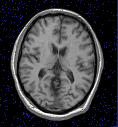 This is an MRI scan. An MRI shows the anatomy of a patient. Dr. Bremner and group
found an 8% decrease in the volume of a structure involved in memory called
the right hippocampus in PTSD victims. Some of the studies that are being
carried out at the Trauma Assesment Unit plan to discover why this alteration
occurs.
This is an MRI scan. An MRI shows the anatomy of a patient. Dr. Bremner and group
found an 8% decrease in the volume of a structure involved in memory called
the right hippocampus in PTSD victims. Some of the studies that are being
carried out at the Trauma Assesment Unit plan to discover why this alteration
occurs.
 A pet scan uses a
radioactive isotope to show areas of brain activity. The brighter sections
indicate elevated regions of activity. By comparing PET scans of individuals
with PTSD to those without PTSD, we can decipher some of the functions
that have been changed by the trauma.
A pet scan uses a
radioactive isotope to show areas of brain activity. The brighter sections
indicate elevated regions of activity. By comparing PET scans of individuals
with PTSD to those without PTSD, we can decipher some of the functions
that have been changed by the trauma.
For more information on imaging technologies look at these sites.
The Imaging center at the Yale Medical School (graphics intensive)
If you have medical concerns about PET scans or questions about imaging in general please send email